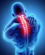
Neck Pain

CASE REPORT CONDUCTED AND PRESENTED BY C GROBBELAAR
April 2015
CONTENTS
Contents Page 1
Abbreviations 2
Abstract 3
Introduction 4
Reporting the Case
- Assessment 7
- Management and outcomes 10
Discussion 12
Conclusion 15
Limitations and recommendations 16
References 17
Appendices 20
- Patient consent form
- Patient assessment and clinical reasoning
ABBREVIATIONS
P1 – primary complaint
P2- secondary complaint
ROM – range of movement
PAIVM – passive accessory intervertebral joint movement
PPIVM – passive physiological intervertebral joint movement
PA – posterior-anterior
OP – overpressure
EOR – end of range
ABSTRACT
Neck pain is a common complaint treated by physiotherapists, and can be caused by biomechanical overload due to sustained poor postures. Combined manual therapy treatments have been shown to be effective in the treatment of mechanical neck pain.
The objective of this study was to show the effect of a combination treatment on a patient with mechanical neck pain, aggravated by sustained poor working postures. The patient presented with limitation of cervical lateral flexion left due to right upper trapezius spasm, and limitation of the movements of lateral flexion and rotation to the right, due to hypomobility of the right C34 zygapophyseal joint. Treatment consisted of specific soft tissue mobilisation, the long lever downslope joint mobilisation technique, both of which had an effect on the upper trapezius spasm. The patient was also taught to correct his posture. The patient’s symptoms were cleared within two treatments.
The effectiveness of this treatment was most likely due to the anatomical links found between the C34 zygapophyseal joint and the upper trapezius muscle. Mechanical neck pain in this patient was effectively managed using a combination of manual therapy techniques, consisting of specific soft tissue mobilisation and the long lever downslope joint mobilisation technique.
INTRODUCTION
Neck pain is a common complaint, for which patients are frequently referred to physiotherapists (De Hertogha et al 2007). At any given time, 19% of the population may be suffering with chronic neck pain, and there is an estimated lifetime prevalence of up to 70% (De Hertogha 2007; Macdermid et al 2009). Neck pain can be classified as mechanical neck pain with a reasonable level of certainty by the following clinical findings on presentation: the patient is younger than 50 years old; they have had acute neck pain for less than 12 weeks; their symptoms are isolated to their neck, and they have restricted cervical range of movement (Childs et al 2009).
Some common problematic activities for patients suffering with mechanical neck pain may involve prolonged poor sitting postures, prolonged, repetitive or strenuous upper limb activities, and movements at the end of range (O’Leary et al 2009). It is hypothesised that spinal posture can have an effect on kinematics, motor control and spinal loading. Altered craniocervical postures, especially chin protraction have been found to alter cervical spine kinematics, thereby altering the load placed on spinal structures, which will ultimately lead to pain (Straker et al 2008). The cervicoscapular muscles (including upper trapezius) may also be affected by prolonged or end of range postures, which, among other things, have been shown to affect cervical muscle performance, and encourage the development of myofascial trigger points (O’Leary et al 2009). These sustained and poor postures are commonly found in many working environments, and may be applicable to many of the patients that are treated by physiotherapists.
Combined interventions of joint mobilisation, soft tissue mobilisation and manual stretching techniques have been found to be effective in the management of both acute and chronic neck pain (Childs et al 2009). Combined interventions have also been shown to be more effective than specific techniques used on their own, as no individual technique has been shown to be superior over another (Hurwitz et al 2008; Gross et al 2009). This improvement in results using combined interventions may in part be due to the anatomical link between the cervicoscapular muscles and the effect that these muscles can have on the somatic and articular structures of the cervical spine, specifically C2, C3 and C4 zygapophyseal joints (Andrade et al 2008). The joint involvement is possibly due to the compressive forces created by the attachments of the cervicoscapular muscles, and the transfer of weight through the cervical spine from the upper extremities (Jull et al 2008, Andrade et al 2008).
There are many different treatment approaches that can be used in the treatment of neck pain, and research has been done into the effectiveness of many of these techniques in the management of specific classifications of neck pain. Evidence has shown that manual mobilisation may be effective in the treatment of younger patients with more acute symptoms, and no signs of nerve root compression (Fritz and Brennan 2007; Hurwitz et al 2008). The long lever downslope technique as described by Hing has been shown to be effective in restoring a loss of rotation or side bending towards the painful side, in patients with mechanical neck pain. These patients show key objective findings of asymmetry, range of motion changes, tissue texture changes and tissue tenderness (Hing 2003).
This case report demonstrates the effective management of a patient presenting with mechanical neck pain, using a combined treatment approach of specific soft tissue mobilisations, long lever downslope joint mobilisation, and postural correction exercises.
REPORTING THE CASE
-ASSESSMENT
The patient, a 35year old male, presented with a 3-day history of right sided middle to lower neck pain and stiffness. He described his primary complaint (P1) as a deep dull ache that was felt constantly while he was awake, but which fluctuated in intensity between a 3/10 and an 8/10 on the numeric pain scale. His secondary complaint (P2) was described as a deep generalised neck stiffness that he felt constantly when he was awake, that was not painful, but he did feel that it restricted his movement.
His symptoms were brought on by four days of long hours in awkward positions at work, as well as long hours driving, and sleeping in different beds. His pain was aggravated to 8/10 after working until one o’clock in the morning, turning his head and looking up under machines, and was eased slightly by a hot shower and Myprodol. He woke with P1 a 3/10 ache in his right lower cervical spine, which worsened to 6/10 by the end of the day. He also felt a general right-sided stiffness and limitation of his neck movement P2. He had no history of trauma or other significant neck problems, and no neurological symptoms.
Observation showed the patient to have a posture of rounded shoulders and a poking chin. His most aggravating movement as demonstrated was cervical flexion, combined with left rotation to the end of range, which increased P1 from 3/10 to 6/10. In this position he also felt P2 stiffness. On return to neutral his symptoms returned to 3/10. Lateral flexion to the right was limited to ¾ range-of-movement (ROM), and reproduced P2 in the right mid cervical spine. Lateral flexion to the left reproduced P1 on the right at ¾ ROM. Rotation to the right was restricted in the mid cervical spine at ¾ ROM, and the restriction felt similar to P2.
When the patient’s scapular position was corrected, he had full active range of cervical rotation to the right when compared with cervical rotation to the left, but still felt the restriction of P2. Muscle length tests showed shortened fibres of upper trapezius bilaterally. Placing the right upper trapezius on stretch reproduced P1. Soft tissue palpation found a taut band in right upper trapezius, and active TrP1 that reproduced P1, and a mild referral into the side of the head behind the eye.
Passive accessory intervertebral movements (PAIVM’s) and passive physiological intervertebral movements (PPIVM’s) as described by Maitland were used to determine the exact level of the joint mobility changes (Maitland 1998). GrIII unilateral posterior-anterior (PA) PAIVM’s on the right C3 C4 vertebrae were painful and stiff, in a similar region to P2. Cervical PPIVM’s found restricted movement at C3,4 level on the right during the individual movements of both passive lateral flexion, and rotation to the right.
After examination the patient’s reported level of pain was 2/10, and he reported that his neck felt looser. Lateral flexion to the left reproduced P1 5/10. Lateral flexion to the right still felt restricted in the mid cervical region, but movement did feel “a little bit easier in general”.
The patient had no yellow flags that were noted. He was not concerned about his neck pain being a serious problem, as he was aware that he had been working long hours in awkward positions, but he was irritated by the continuing discomfort, and did not want it to limit his sporting activity during the following few days.
MANAGEMENT AND OUTCOMES
Treatment consisted of soft tissue mobilisations, specifically myofascial stretching and trigger point release to the right upper trapezius, after which re-assessment found P1 at rest to be 0/10, and 3/10 with lateral flexion to the left. P2 still limited rotation to the right and lateral flexion to the right to 3/4ROM. Long lever downslopes were selected as the treatment of choice based on criteria as described by Hing. These criteria were that the patient had only two directions of movement loss that were biomechanically linked (cervical lateral flexion right and rotation right) for his symptom of P2. The patient’s symptoms were also biomechanical in nature with a firm joint end feel, and a regular recognisable biomechanical pattern (Hing 2003). Long lever downslope mobilisations were then performed on C34 on the right, working just into the start of resistance, 2x30s. After this, P1 at rest was 0/10, and lateral flexion to the left was a 1/10, felt more as a mild pull than pain. He felt a mild stiffness with lateral flexion and rotation to the right, but this was not localised to the mid cervical region.
With subjective examination at his second treatment 6 days later, the patient reported not having felt P1 since his last treatment, but was still feeling a mild neck stiffness. During the objective re-examination, lateral flexion to the left had full pain free range, with overpressure (OP). During both individual movements of lateral flexion and rotation to the right he felt a slight stiffness on the right, but it was not specifically identified as P1 or P2.
As the patient’s signs and symptoms had improved, joint mobilisation using the long lever downslope technique at C34 on the right was progressed into end of range (EOR) resistance, 2x30s. On re-assessment of lateral flexion and rotation to the right, he had full ROM with overpressure.
The patient was then educated as to how to correct his sitting posture. During initial assessment, the patient’s ROM of cervical rotation had been found to be limited towards the right to ¾ ROM, limited by P2. When the therapist corrected the patient’s posture by assisting with the positioning of their scapula in neutral, and cervical rotation ROM was re-assessed, the active range of movement was comparable to the full pain free range found with rotation to the left, although there was still a slight discomfort of P2 felt at the end of active range of rotation right. This procedure was used to demonstrate to the patient the effect of improved sitting posture on their symptoms.
As a continuation of his second treatment, the physiotherapist assisted the patient into a correct sitting posture, by facilitating the correct lumbar spinal curvature, slight chin retraction, and setting the scapulae in the correct position. The patient then held this position for 5 seconds. This positioning was facilitated a total of 3 times, then the patient was asked to find this position themselves, and hold for 5 seconds. This unassisted repositioning was performed 3 times. The patient was advised to gradually increase the number and duration of holds to 10x10seconds over the next 10days, and advised to assume this posture as often as possible when doing computer work and driving. He was also advised to check his pillow heights in order to keep his head and neck in neutral, and ensure that his neck is well supported when sleeping.
DISCUSSION
This patient’s symptoms were eased by a combined treatment consisting of specific soft tissue mobilisations and the long lever downslope technique to mobilise his hypomobile right C34 zygapophyseal joint. His primary complaint (P1) of right sided lower neck pain was eased by soft tissue mobilisation and trigger point release of his right upper trapezius muscle. It was relieved from a 2/10 to 0/10 at rest, and 5/10 to 3/10 when retesting his comparable sign of lateral flexion to the left, which placed his right upper trapezius on a stretch. This trigger point had probably been activated originally by the patient’s sustained neck position looking up under machines, as trigger points in the fibres of upper trapezius can be activated when the head and neck are almost fully actively rotated towards the other side (Travell and Simons 1993). The most aggravating movement as demonstrated by the patient was a combined movement of rotation left and flexion. When demonstrating his neck position as if he were looking up under a machine, he also introduced an additional movement of lateral flexion to the right. These combined movements would place his right upper trapezius in a maximally shortened position, which could potentially activate trigger points in this muscle.
Travell has also shown that trigger points in upper trapezius may be perpetuated by asymmetry of the upper limbs and shoulder girdle. The weightbearing function of the upper trapezius in supporting the upper limb can be over stressed when the arms are unsupported for an extended period of time, or through prolonged elevation of the shoulders as an expression of anxiety or stress (Travell and Simons 1993). Occupational factors have also been shown to play a large contributing role in the development of neck pain (Gross et al 2009). When the position of looking up under machines was held for an extended period of time during the course of his working day, in combination with the other potentially aggravating activities of a long stressful working day, driving long distances, and sleeping in different beds with unfamiliar pillows, these activities in combination would be likely to activate a trigger point in his upper trapezius muscle.
Hypomobility of the right C34 zygapophyseal joint was also found on palpation examination, and this was similar to the predominantly right-sided neck stiffness (P2) of which the patient complained. PAIVM’s and PPIVM’s as described by Maitland were used to determine the exact level of joint mobility changes (Maitland 1998). The patient’s symptoms of P2 were reproduced with GrIII unilateral PA PAIVM’s of the right C34 zygapophyseal joint. The downslope technique as described by Hing et al was chosen to mobilise this joint as it is a side bending and extension mobilisation, and can be used to restore a loss of extension, side bending or rotation towards the side of movement (Hing et al 2003). As the patient was relatively young, with no history of underlying cervical pathology, and stiffness being more noted in this joint than pain, a long lever technique was chosen.
When the patient was retested after joint mobilisation to determine the effectiveness of the long lever downslope mobilisation technique of his right C34 zygapophyseal joint for treatment of P2, he felt not only an improvement of P2, but he also felt further relief of his P1 symptoms. This is comparable to the correlation that Travell and Simons found between somatic or articular dysfunction of the C2, 3 and 4 vertebrae, and trigger points in the upper trapezius, which often co-exist. C4 joint involvement can cause a referral pain over the area of the upper trapezius. This can cause secondary involvement of the upper trapezius muscle, and the muscle may become hyperirritable, which can lead to the development of trigger points (Travell and Simons 1993).
The cervicoscapular muscles are also thought to have a mechanical impact on the cervical spine. Although there is still some speculation about this matter, there is a likelihood of compressive forces being placed on the upper cervical motion segments due to the superior attachments of the upper trapezius and levator scapulae muscles, and the associated transfer of weight from the upper extremities (Jull 2008, Andrade 2008). During the initial physical assessment, the patient’s range of movement with rotation to the right improved from ¾ to full active range with correction of his scapular position. A study by Andrade et al showed that elevation of the scapulae by passively supporting the upper limbs resulted in an increase in the range of cervical rotation (Andrade et al 2008). Although the exact mechanism for this increase in range, which occurs in both healthy, painfree individuals, and those with cervical dysfunction, is unknown (Andrade et al 2008), there is a possibility that it occurs as a result of decreased tension in the aforementioned cervicoscapular muscles, and subsequent decompression of the cervical zygapophyseal joints.
CONCLUSION
A combination of manual therapy techniques was effective in reducing pain and improving range of movement in mechanical neck pain in this patient. The improvement in this patient’s pain is hypothesized to be largely due to the biomechanical interaction between the joints and muscular attachments, and underlying postural loading mechanisms, as joint mobilisation was also responsible for relieving muscle spasm. All of these factors need to be addressed in order to ensure effective management of this patient.
LIMITATIONS AND RECOMMENDATIONS
The patient was taught scapular positioning exercises and chin tuck in order to correct his posture in sitting as part of his second treatment, and these were also given as home exercises. There was however no further follow-up treatment in order to assess the effect of, and progress these exercises. This can be seen as a limitation of this study. There is also currently very little published research showing the efficacy of the long lever downslope joint mobilisation. This could be a researched in further studies.
REFERENCES
Andrade, GT; Azevedo, DC; Lorentz,ID; Neto, RSG; Do Pinho, VS;McDonnell, MK, Van Dillen, LR; 2008. Influence of scapular position on cervical rotation range of motion. Journal of Orthopaedic and Sports Physical Therapy 38:11.
Childs, JD; Cleland, JA; Elliott, JM; Teyhen, DS; Wainner, RS; Whitman,JM; Sopky, JM; Godges, JJ; Flynn, TW; 2008. Neck Pain: Clinical practice guidelines linked to the international classification of functioning, disability, and health from the orthopaedic section of the American Physical Therapy Association. Journal of Orthopaedic Sports Physical Therapy38(9):A1-A34.
De Hertogha, WJ; Vaesa, PH; Vijverman, V; De Cord, A; Duqueta, W; 2007.
The clinical examination of neck pain patients: The validity of a group of tests
Manual Therapy 12: 50–55.
Fritz, JM; Brennan,GP; 2007. Preliminary examination of a proposed treatment-based classification system for patients receiving physical therapy interventions for neck pain. Physical Therapy 87:513–524
Gross, AR; Haines, T; Goldsmith, CH; Santaguida, L; McLaughlin, LM; Peloso, P; Burnie, S; Hoving, J; Cervical Overview Group (COG); 2009. Knowledge to action: A challenge for neck pain treatment. Journal of Orthopaedic & Sports Physical Therapy 39 : 5.
Hing, WA; Reid, DA; Monaghan, M; 2003. Manipulation of the cervical spine.
Manual Therapy 8(1), 2–9
Hurwitz, EL; Carragee, EJ; van der Velde,G; Carroll, LJ; Nordin, M; Guzman, J; Peloso, PM; Holm, LW; Cote, P; Hogg-Johnson, Sheilah; Cassidy, JD; Halderman, S; 2008. Treatment of Neck Pain: Noninvasive Interventions
Results of the Bone and Joint Decade 2000–2010 Task Force on Neck Pain and Its Associated Disorders. SPINE Volume 33:4; 123–152
Jull, G; Sterling, M; Falla,D; Treleaven, J; O’Leary, S; 2008. Whiplash, headache, and neck pain: Research-based directions for physical therapists. www.elsevierhealth.com :Churchill Livingstone. Elsevier.
Macdermid, JC; Walton, DM; Avery, S; Blanchard, A; Etruw, E; McAlpine, C; Goldsmith, CH; 2009. Measurement properties of the neck disability index: A systematic review. Journal of Orthopaedic & Sports Physical Therapy 39:5.
Maitland, GD; 1998. Vertebral Manipulation: Fifth edition. Linacre House,Jordan Hill, Oxford OX28DP: Butterworth-Heinemann.
O’Leary, S; Falla, D; Elliott, JM; Jull, G, 2009. Muscle dysfunction in cervical spine pain: Implications for assessment and management. Journal of Orthopaedic & Sports Physical Therapy 39:5.
Straker, LM; O’Sullivan, PB; Smith, AJ; Perry, MC; Coleman, J; 2008. Sitting spinal posture in adolescents differs between genders, but is not clearly related to neck/shoulder pain: an observational study. Australian Journal of Physiotherapy 54: 127-133.
Travell, JG; Simons, DG.1993. Myofascial pain and dysfunction: The trigger point manual volume 2. 530 Walnut Street, Philadelphia, PA 19106: Lippincott Williams and Wilkins
APPENDICES
CONSENT FORM FOR CASE REPORTS[1]
Explanation to the patient:
Each candidate is required to submit a report on a patient that they have assessed and treated in clinical practice.
The purpose of the case report is for the candidates to explain their thinking and reasoning processes against the background of applicable research.
Patient consent for publication of information about them in a case report for certification purposes and possibly in an academic journal:
Name of person described in the report:__________________________
Subject matter of the report:__________________________
Title of case report:_________________________________________________
Name of Physiotherapist ____________________
I_________________________________________ [insert full name of patient] give my consent for this information about MYSELF OR MY CHILD OR WARD/MY RELATIVE [insert full name]:_________________________, relating to the subject matter above (“the Information”) to appear in a case report, or to be used for the purpose of an academic journal publication.
I understand the following:
- The Information will be published without my name/child’s name/relatives name attached and every attempt will be made to ensure anonymity. I understand, however, that complete anonymity cannot be guaranteed, due to the nature of the assessment.
- The Information may be published in a case report or journal which is read worldwide or an online journal. Journals are aimed mainly at health care professionals but may be seen by many non-doctors, including journalists.
- The Information may be placed on a website.
- I can withdraw my consent at any time before online publication, but once the Information has been committed to publication it will not be possible to withdraw the consent.
Signed:__________________________________ Date: ______________________
Signature of requesting medical practitioner/health care worker:
_____________________Date:______________
[1] Adapted from BMJ Case Reports consent form.


